Management of double vision
Double vision, medically known as diplopia, is a visual condition where a person sees two images of a single object. These images may appear side by side (horizontal diplopia), one above the other (vertical diplopia), or diagonally displaced.
Diplopia can be temporary or permanent, and its management depends on identifying and treating the underlying cause. In some cases, it may signal a minor eye muscle imbalance, while in others, it could be an early warning sign of a neurological or systemic disorder.
Types of Double Vision
Understanding the type of diplopia helps guide diagnosis and management.
Monocular Diplopia
Persists when one eye is open and disappears when that eye is closed. Usually due to an issue within the eye itself (e.g., refractive error, cataract, corneal irregularity).
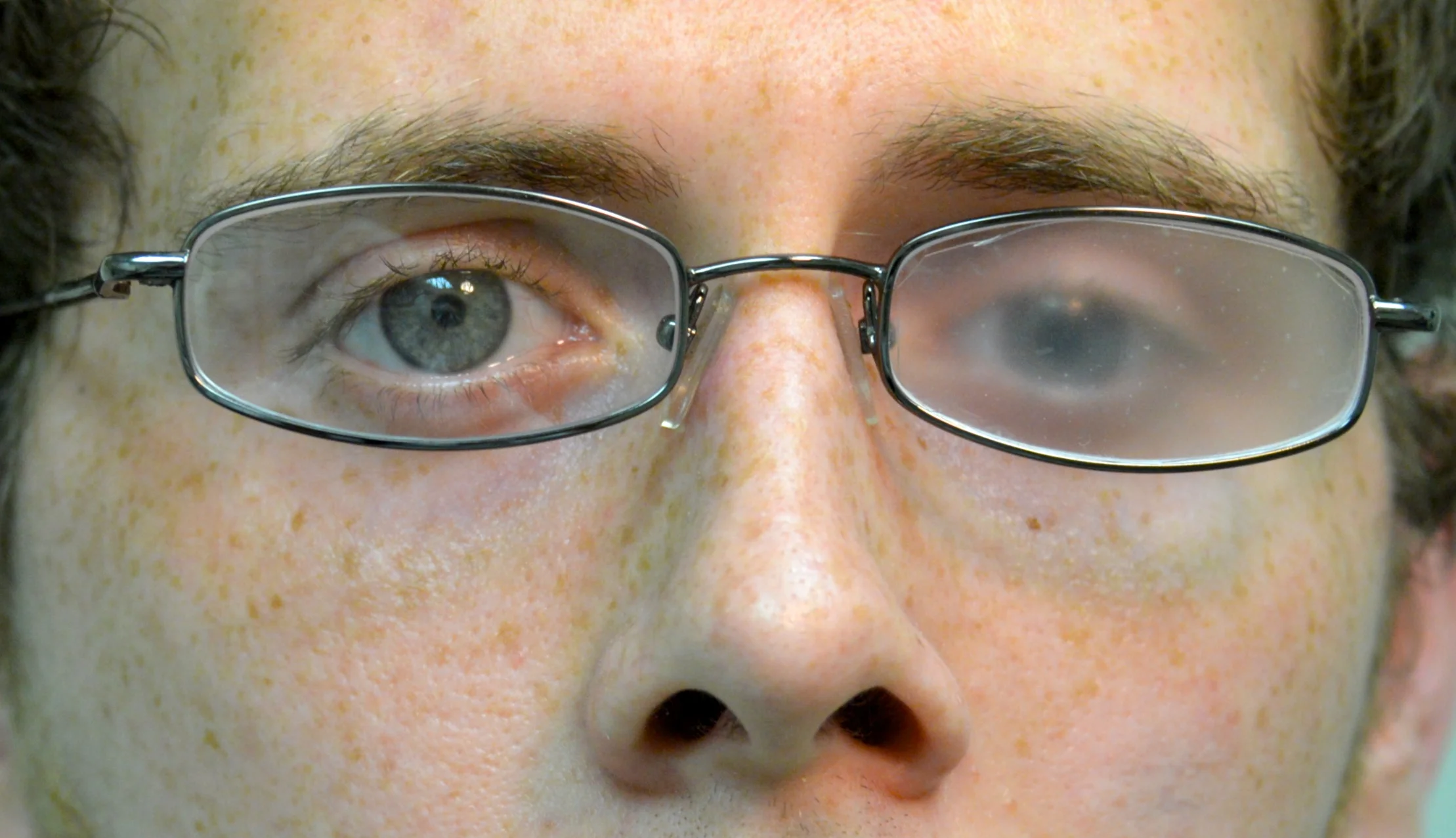
Binocular Diplopia
Present only when both eyes are open and resolves when one eye is closed. Typically results from misalignment of the eyes due to muscle, nerve, or brain-related causes.
Common Causes of Double Vision
Monocular Causes
- Refractive errors (e.g., astigmatism)
- Corneal irregularities (e.g., keratoconus, corneal scarring)
- Lens problems (e.g., cataract or lens dislocation)
- Retinal abnormalities
Binocular Causes
- Cranial nerve palsies (III, IV, VI): Affect eye muscle movement
- Thyroid eye disease: Causes muscle swelling and restricted movement
- Myasthenia gravis: Leads to fluctuating weakness of eye muscles
- Orbital trauma or fracture: Can trap muscles, preventing normal movement
- Brainstem lesions or stroke: Affect eye coordination and movement control
- Diabetes mellitus: Can cause nerve palsy leading to temporary diplopia
Symptoms Associated with Double Vision
- Seeing two overlapping or separate images
- Eye strain or discomfort
- Headache
- Nausea or dizziness
- Difficulty reading or focusing
- Tilting or turning the head to compensate for misalignment
Diagnosis of Double Vision
Proper diagnosis requires a detailed clinical evaluation and diagnostic testing to identify the cause.
Comprehensive Eye Examination
- Assess vision in each eye separately and together
- Evaluate ocular alignment and movement
Neurological Evaluation
To rule out cranial nerve palsy, brain injury, or systemic neurological disorders
Imaging Tests
MRI or CT scan: To detect tumors, aneurysms, or structural abnormalities in the brain or orbit
Blood Tests
To check for underlying systemic diseases (e.g., diabetes, thyroid disorders, myasthenia gravis)
Specialized Tests
Hess chart or synoptophore to measure muscle deviation and eye coordination
Management of Double Vision
Treatment of double vision depends on its type, cause, and duration. The goal is to restore single vision, relieve discomfort, and prevent further complications.
1. Treating the Underlying Cause
- Refractive errors: Corrected with glasses or contact lenses.
- Cataracts: Surgical removal often resolves monocular diplopia.
- Thyroid eye disease: Managed with medications, corticosteroids, or orbital decompression surgery.
- Myasthenia gravis: Treated with anticholinesterase drugs, immunosuppressants, or thymectomy.
- Diabetic nerve palsy: Usually resolves spontaneously with good blood sugar control.
2. Non-Surgical Management
-
Prism Glasses
Prisms bend light to realign the two images into one. Often used for small, stable deviations. Available as temporary stick-on Fresnel prisms or ground-in prisms in permanent glasses.
-
Occlusion Therapy
Eye patching or opaque contact lenses can eliminate double vision in one eye. Often a temporary measure, especially when recovery is expected.
-
Vision Therapy / Orthoptic Exercises
Exercises that strengthen eye muscles and improve coordination. Beneficial for cases like convergence insufficiency or mild muscle imbalance.
-
Botulinum Toxin (Botox) Injections
Temporarily weakens overacting eye muscles to help realign the eyes. Commonly used for acute nerve palsy or when surgery is not yet indicated.
3. Surgical Management
If double vision persists and is stable for several months, strabismus (eye muscle) surgery may be considered. The surgeon adjusts the position or length of extraocular muscles to achieve better alignment. Often performed under local or general anesthesia. Adjustable sutures may be used to fine-tune alignment after surgery. Post-surgery, patients may still need minor prism correction or visual therapy for optimal results.
Prognosis
Transient diplopia (such as from diabetic nerve palsy or minor trauma) often resolves within weeks to months. Chronic or neurological causes may require long-term management with prisms or surgery. Early diagnosis and treatment greatly improve visual outcomes and quality of life.
When to Seek Immediate Medical Attention
Double vision can sometimes be a sign of a serious neurological problem. Seek urgent medical care if diplopia appears suddenly or is accompanied by:
- Severe headache
- Difficulty speaking or swallowing
- Weakness or numbness in limbs or face
- Sudden vision loss